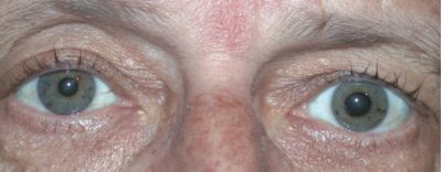
An 18 year old man presents to the emergency department after being struck in the right eye with a baseball. He reports pain, photophobia, and monocular diplopia. Visual acuity is 20/40 in the right eye and 20/20 in the left. Intraocular pressure is 10 mmHg in the right eye and 16 mmHg in the left. Slit lamp examination of the right eye reveals a formed anterior chamber and a peripheral iris defect with a second pupil-like opening near the inferotemporal limbus. There is mild layered hyphema and iridodonesis. Dilated fundus examination is unremarkable.
Which of the following is the most appropriate next step in management of this patient’s iris finding?
A. Immediate surgical repair of the iris root
B. Observation with topical corticosteroids and cycloplegics
C. Emergent trabeculectomy
D. Laser peripheral iridotomy
E. Intravitreal anti VEGF injection
Correct Answer: B. Observation with topical corticosteroids and cycloplegics
Explanation:
This patient has traumatic iridodialysis, a disinsertion of the iris root from the ciliary body, commonly caused by blunt ocular trauma. Classic findings include a peripheral iris defect, corectopia, monocular diplopia, glare, photophobia, and sometimes hyphema. Intraocular pressure may initially be low due to ciliary body shock.
Small or minimally symptomatic iridodialyses are typically managed conservatively with topical corticosteroids to control inflammation and cycloplegics to reduce pain and prevent synechiae. Surgical repair is reserved for large defects causing significant glare, diplopia, cosmetic concerns, or uncontrolled intraocular pressure. Trabeculectomy and laser peripheral iridotomy are not indicated in this setting, and anti VEGF therapy has no role in acute traumatic iridodialysis.
Patients should also be monitored for associated injuries such as angle recession, traumatic cataract, retinal tears, and secondary glaucoma on follow up.